TGen's Center for Spatial Multi-Omics (COSMO)  Header image

TGen's Center for Spatial Multi-Omics (COSMO)

Mapping your samples, one molecule at a time!

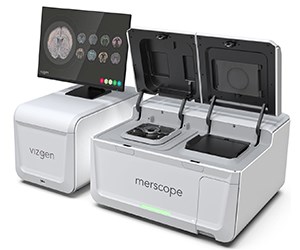
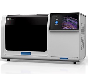
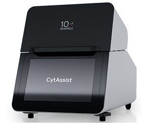
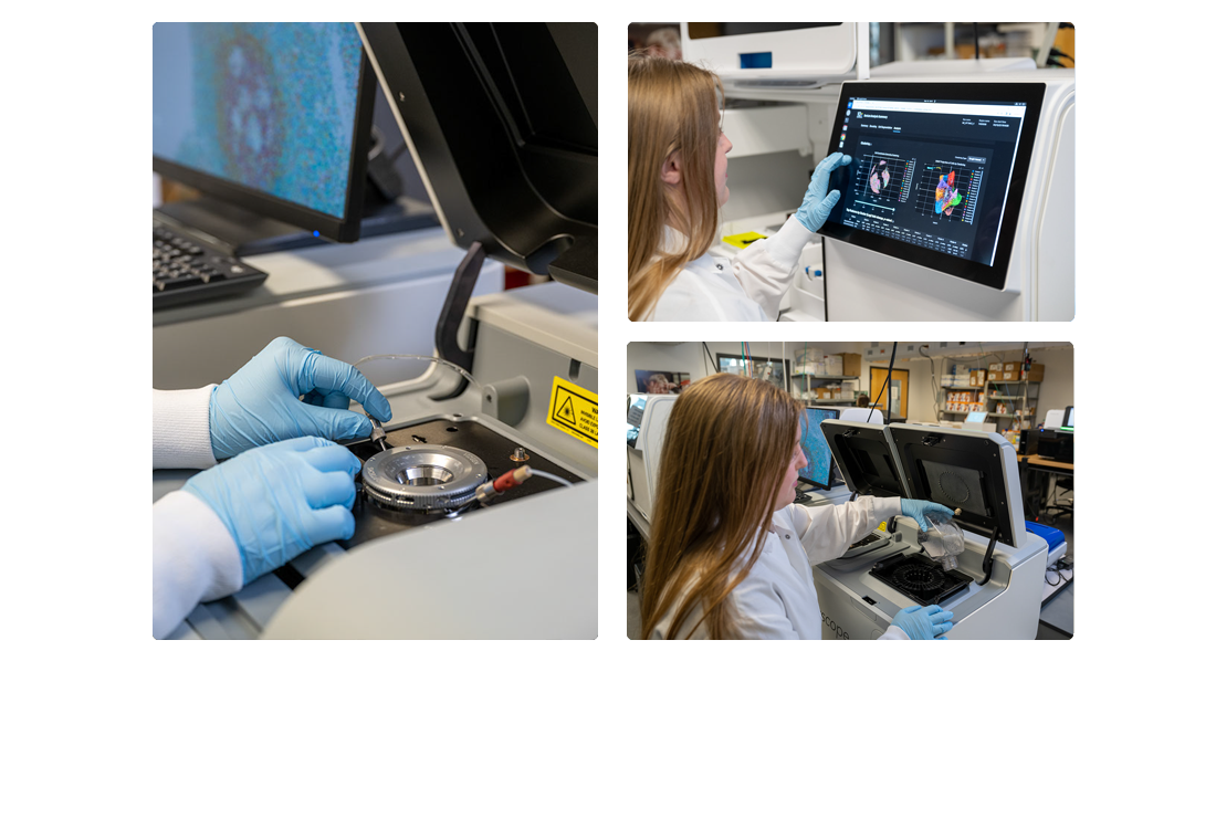
TGen always strives to be at the forefront of new research technologies. At the TGen's Center for Spatial Multi-Omics (COSMO), we want to help you dive into the world of spatial biology! By offering a variety of services and instrument options, we allow you to visualize your samples in brand new ways. With spatial transcriptomics, you can profile the expression of a large tissue at sub-cellular RNA resolution. Investigate individual cells while preserving cellular niches and anatomical structures for histological analyses.


Available Instruments & Services

Interested in adding spatial to your project? Contact us! [email protected]

Vizgen MERSCOPE Ultra


Custom Panels up to 960 Genes
Imageable area up to 3cm2
Cell Segmentation
Protein Stain Add-ons 

Using Multiplex Error-Robust Fluorescence In Situ Hybridization (MERFISH), the MERSCOPE Ultra allows the spatial mapping of 960 custom RNA targets at subcellular resolution (≤100nm resolution). 

MERSCOPE Ultra has two different sized flowcells to allow for experimental customization with varying sizes of tissue: 1.25cm2 and 3cm2

On-instrument cell segmentation uses machine learning to combine nuclei staining, total mRNA abundance (poly-A), and cell boundary staining. 

Predesigned panels available:

Human PanCancer Pathways, Human Immuno-Oncology, Mouse PanNeuro 

Compatible with both frozen and FFPE samples.

10X Genomics Xenium


Custom Panels up to 480 Genes
Imageable area of 2.88cm2
Cell Segmentation
Post-Run H&E or IF 

Using high-throughput RNA Fluorescence In Situ Hybridizaiton, the Xenium Analyzer allows the spatial mapping of 480 custom RNA targets, or 5000 predesigned RNA targets at subcellular resolution (~0.2 microns/pixel). 

Each instrument run profiles two slides for a total imageable area of 5.76cm2 per run. 

On-instrument cell segmentation uses machine learning to combine nuclei staining, interior-RNA abundance, interior-protein abundance, and cell boundary staining.

 H&E or IF staining can be performed on the same tissue slide following a Xenium run, allowing for the unique overlap of pathological analysis and gene expression. 

Predesigned panels available:

Human Brain, Human Breast, Human Lung, Human Immuno-Oncology, Human Colon, Human Skin, Human Multi-Tissue, Human 5000 Gene, Mouse Brain, Mouse Multi-Tissue, Mouse 5000 Gene 

Compatible with both frozen and FFPE samples.

10X Genomics Visium HD


Whole Transcriptome
Imageable area of 0.85cm2
FFPE Blocks or Archived Slides
H&E or IF Image Mapping 

Using probe-based capture of transcriptomic data, Visium HD allows whole transcriptome level mapping of a tissue section in 2x2 µm bins with no gaps in tissue coverage. These probes can then be sequenced and mapped back to their bin location on the tissue slide image. 

Constructed gene expression library is sequenced at a recommended depth of 275 million read pairs for capture areas fully covered by tissue. 

Sample slides can be H&E or IF stained prior to Visium HD run. 

Compatible with FFPE blocks or archived FFPE slides.

 TGen's Center for Spatial Multi-Omics (COSMO)  Donation image

TMA Construction

For any of our spatial multiomic technologies, we can offer the construction of a compatible TMA for FFPE tissues. This allows us to fit multiple samples within a single capture area, thus decreasing your run cost per sample.

TMAs can be constructed with 3mm, 4mm, or 5mm cores punched from FFPE blocks. Depending on size chosen, this equates to:

4-23 samples per capture area on MERSCOPE
7-17 samples per capture area on Xenium
Up to 4 samples (3mm) per capture area on Visium HD

MEET THE TEAM

Nicholas Banovich
Nicholas Banovich
COSMO Director, Associate Professor
Emerging Technologies and Genome Science
Arianna Williams
Arianna Williams
COSMO Research Associate
TGen
Evan Mee
Evan Mee
COSMO Bioinformatician
TGen